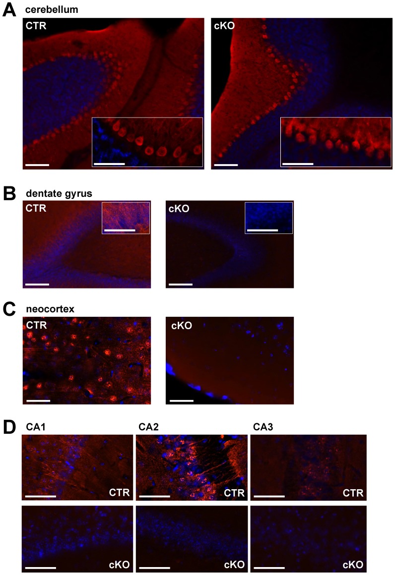
Figure 1. Immunohistochemical comparison of the expression pattern of CaV2.1 in the forebrain and cerebellum of CTR and cKO mice shows a selective knock-out in the hippocampus and neocortex of cKO mice.
(A) CaV2.1 immunofluorescence staining in cerebellum demonstrating a high expression of CaV2.1 in Purkinje cell layers of both CTR and cKO mice indicating that the NEX/Cre-mediated knock out strategy did not affect CaV2.1 expression in cerebellum. (B) to (D) CaV2.1 immunofluorescence staining of horizontal sections of the dentate gyrus, neocortex and hippocampal CA1 to CA3 regions demonstrates expression of CaV2.1 throughout CA1, CA2, CA3, dentate gyrus and neocortex in CTR and a hardly detectable expression of CaV2.1 in cKO mice. CaV2.1 expression is shown in red, DAPI in blue. Scale bars: (A) 100 µm (inset 50 µm); (B) 100 µm (inset 50 µm); (C) 50 µm; (D) 50 µm.

