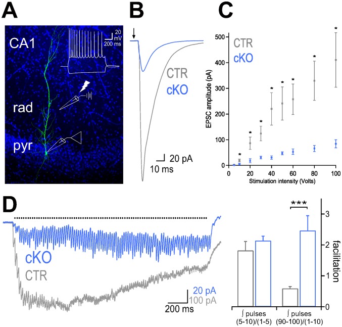
Figure 2. Synaptic transmission at Schaffer collateral-mediated inputs onto CA1 pyramidal cells of cKO mice is impaired.
(A) Confocal image stack of an intracellular labeled CA1 pyramidal cell (PC) in a cKO mouse counterstained with DAPI. Inset, superimposed voltage traces of the same PC in response to a 100 pA depolarizing and 50 pA hyperpolarizing current injection (1 s). (B) Representative traces of pyramidal cell EPSCs evoked by extracellular stimulation of Schaffer collaterals (stimulation intensity 100 volts), in CTR (gray) and cKO mice (blue). Arrow marks the time point of stimulation. (C) Summary plot of amplitudes of EPSCs evoked by Schaffer collaterals stimulation at different intensities in CTR (n = 7) and cKO (n = 10) PCs. Means + SEM are shown. (D) Left, average of EPSCs evoked by a train of 100 stimulation pulses at 50 Hz (Black dots indicate the time points of extracellular stimulation, stimulation artifacts have been removed for clarity). Right, synaptic charge transferred during trains of Schaffer collateral-mediated EPSCs is characterized by an early facilitation in both CTR and cKO animals, but strong multiple-pulse facilitation in cKO PCs (n = 9) and multiple-pulse depression in CTR PCs (n = 6) of mice. * P≤0.05; ** P≤0.01; *** P≤0.001 (Two-tailed student t-test)

