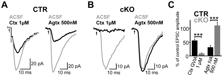
Figure 3. Expression of functional CaV2.1 channels is strongly reduced in the hippocampus of cKO mice.
(A and B) Representative EPSCs recorded in CA1 PCs evoked by extracellular stimulation of the stratum radiatum before (gray) and after (black) bath-application of ω-conotoxin GVIA (left) or ω -agatoxin IVA (right). PC recordings were obtained from CTR (A) and cKOs (B). (C) Bar graph summarizes the residual peak amplitude of EPSCs after toxin application for CTR (each 6 experiments) and cKO (5 vs. 6 experiments) mice. Means + SEM are shown. *** P≤0.001 (Two-tailed Student’s t-test)

