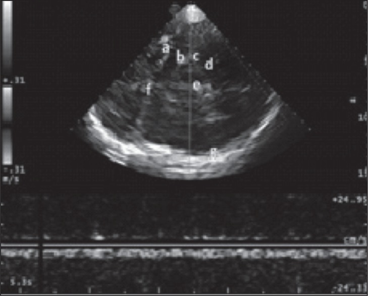
Figure 1 .
Transtemporal approach at the level of midbrain (e) contralateral skull (g). The ultrasonic beam makes it possible to identify: the middle cerebral artery (a) and the contralateral skull (f), the posterior cerebral artery, pre-peduncular part (b), the Rosenthal vein (c) and the posterior cerebral artery, post-peduncular part (d).

