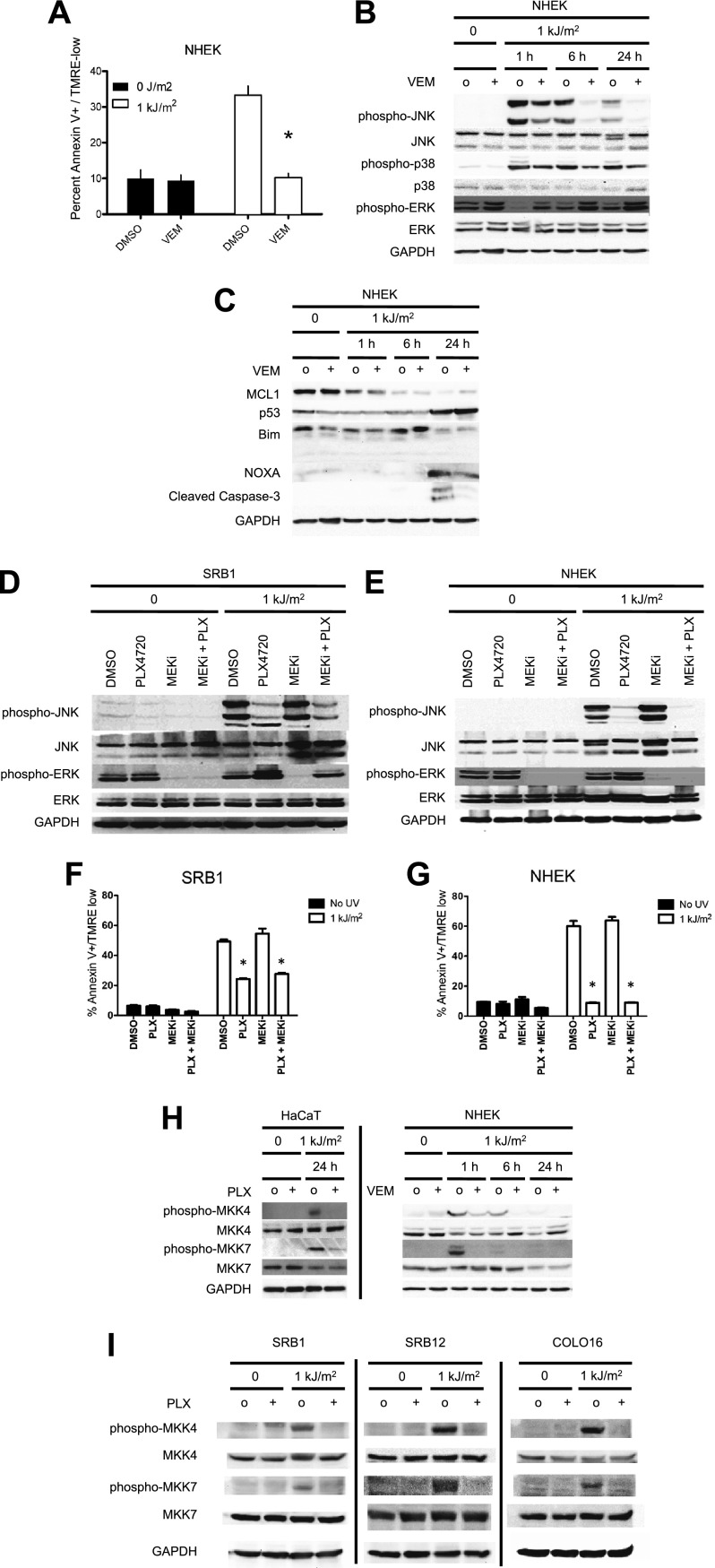
Figure 2. Vemurafenib and PLX4720 suppress apoptosis and JNK signaling in primary human keratinocytes and cSCC cells independently of MEK/ERK signaling.
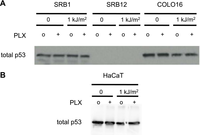
Normal human epidermal keratinocytes (NHEKs) were irradiated with 1 kJ/m2 of UVB in the absence (‘o’, 1:2000 DMSO) or presence (‘+’) of 1 μM vemurafenib and isolated for FACS analysis and protein extracts 24 hr later. (A) Apoptosis was significantly suppressed (70%) in the presence of vemurafenib as measured by FACS for Annexin V+, TMRE-low cells (n = 6, ‘*’ denotes statistical significance at p<0.05). (B) Western blot analysis showed induction of phospho-JNK and phospho-p38 within 1 hr following irradiation, which persisted for at least 24 hr and which was suppressed by vemurafenib at all time points. (C) MCL1 and BIM expression was not significantly modulated by vemurafenib; however, NOXA induction, which occurred at 24 hr, was reduced by vemurafenib. In these primary cells, p53 protein was stabilized by 24 hr and vemurafenib did not affect overall levels. Suppression of apoptosis, as measured by cleaved caspase-3 levels, was observed in the presence of vemurafenib-treated irradiated cells, consistent with the FACS results. To test the relevance of MEK signaling, cSCC (SRB1) and NHEK cells were irradiated with 1 kJ/m2 of UVB in the absence (‘o’, 1:2000 DMSO) or presence of 1 μM PLX4720 singly or in combination with 0.6 μM (NHEK) or 1.2 μM (SRB1) AZD6244 (MEKi) and isolated for FACS analysis and protein extracts 24 hr later. (D) SRB1 and (E) NHEK cells showed induction of phospho-JNK at 24 hr following irradiation, by Western in the presence (lane 7) and absence (lane 5) of MEKi. The addition of MEKi to PLX4720 did not affect the suppression of JNK activation (compare lanes 6, 8) despite potent suppression of phospho-ERK. (F) SRB1 and (G) NHEK cells exhibited a strong suppression of UV-induced apoptosis by PLX4720 (Annexin V+, TMRE-low cells; n = 6, ‘*’ denotes statistical significance at p<0.05) that was likewise unaffected by the addition of MEKi. To test whether upstream kinases in the JNK pathway were inhibited, MKK4 and MKK7 activation was probed in cells. (H) Both phospho-MKK4 and phospho-MKK7 were induced in HaCaT and NHEK cells following irradiation, and this was suppressed in the presence of 1 μM PLX4720 and vemurafenib, respectively. (I) In all cSCC cell lines, SRB1, SRB12, COLO16, phospho-MKK4 and phospho-MKK7 are strongly induced following irradiation, and this is suppressed in all lines by 1 μM PLX4720.


