. 2013 Nov 5;2:e00969. doi: 10.7554/eLife.00969

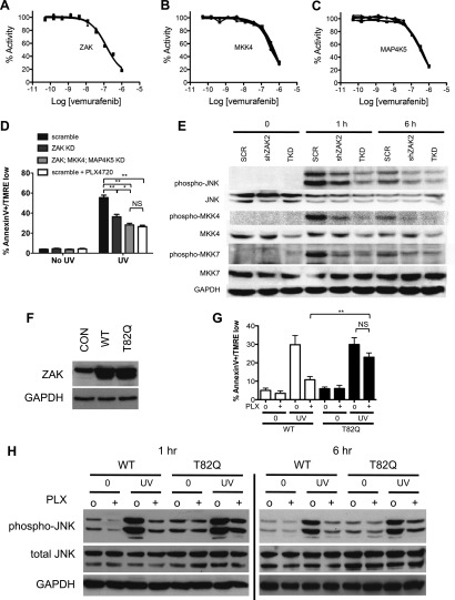
Figure 3. PLX4720 and vemurafenib suppress apoptosis and JNK signaling through inhibition of off-target kinases.

(A–C) In-vitro kinase assays for ZAK, MKK4, and MAP4K5 were performed across a 10-point concentration range from 0.05 to 1000 nM in triplicate, revealing significant inhibition of kinase activity within the nM range for vemurafenib. (D) Lentiviral shRNA knockdown of ZAK singly or in combination with MKK4 and MAP4K5 (triple knockdown, ‘TKD’) was performed revealing potent suppression of apoptosis as measured by FACS for Annexin V+, TMRE-low cells (n = 5, ‘*’ denotes statistical significance at p<0.05, ‘**’ at p<0.01, ‘NS’ is not significant) at 24 hr following single dose UVB irradiation at 720 J/m2. ZAK knockdown and triple knockdown cells exhibit 70% and 94% suppression of apoptosis, respectively, relative to PLX4720-treated cells expressing a non-suppressing shRNA control (scramble, ‘SCR’). (E) Western blots of lysates obtained at 1 and 6 hr post-UV irradiation show potent induction of phospho-MKK4, phospho-MKK7, and phospho-JNK which are all suppressed with progressively increasing effect in ZAK single knockdown (‘shZAK2’) and triple knockdown (‘TKD’) HaCaT cells. (F) Western blots of HaCaT cells electroporated with pcDNA3-wild-type (WT) ZAK and the gatekeeper mutant pcDNA3-(T82Q) ZAK show equivalent expression. (G) HaCaT cells overexpressing ZAK (WT) and ZAK (T82Q) were irradiated with a single dose of UVB irradiation at 720 J/m2 in the absence (‘o’) and presence (‘+’) of 1 μM PLX4720 and apoptosis measured by FACS for Annexin V+, TMRE-low cells (n = 4, ‘**’ at p<0.01, ‘NS’ is not significant) at 24 hr. ZAK (WT) cells are sensitive to PLX4720-mediated suppression of apoptosis (bar 3 vs 4), but drug-treated ZAK (T82Q)-expressing cells undergo significantly more apoptosis than drug-treated ZAK (WT) cells (bar 4 vs 8), with bypass of PLX4720-induced suppression as compared to drug-treated ZAK (WT) cells (paired t-test, p=0.005). (H) Western blots of ZAK (WT) and ZAK (T82Q)-expressing HaCaT cells at 1 hr and 6 hr post-irradiation show that phospho-JNK activation is intact in both cell lines in the absence of drug (lanes 3, 7), but that drug-treated ZAK (T82Q)-expressing HaCaT cells have significantly more phospho-JNK activation at both 1 and 6 hr post-irradiation, as compared to drug-treated ZAK (WT)-expressing cells (lane 4 vs 8).

DOI: http://dx.doi.org/10.7554/eLife.00969.013

Figure 3.

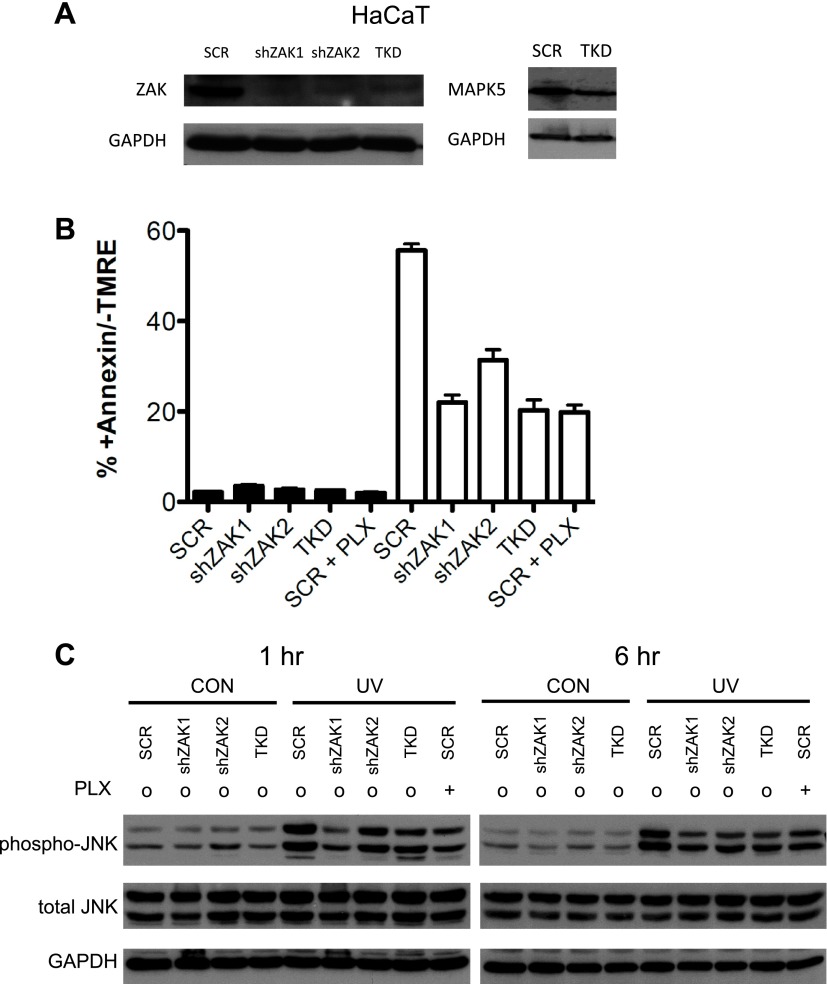
Figure 3—figure supplement 1. Knockdown of ZAK potently inhibits JNK activation and UV-induced apoptosis.

Figure 3—figure supplement 1.

(A) Western blot of HaCaT cells expressing two shRNA clones and HaCaT TKD cells (containing shZAK2) all show significant knockdown of ZAK protein, though shZAK2 produces slightly less knockdown. Approximately 54.6% knockdown (ImageJ) of MAP4K5 is observed in TKD cells. (B) HaCaT cells, expressing non-silencing scramble-shRNA (‘SCR’), shZAK1, shZAK2, or TKD, were either unirradiated (black bars) or irradiated (open bars) with 1 kJ/m2 of UVB in the absence (‘o’, 1:2000 DMSO) or presence (‘+’) of 1 μM PLX4720 and analyzed by FACS for apoptosis (Annexin V+, TMRE-) at 24 hr. UV-induced apoptosis is significantly suppressed by both ZAK shRNA clones in HaCaT cells and in TKD cells. The shZAK2 clone, which results in less knockdown than shZAK1, produces correspondingly less suppression of UV-induced apoptosis (93.7% for shZAK1 vs 67.8% for shZAK2). shZAK1-expressing HaCaT cells, TKD cells, and PLX4720-treated HaCaT scrambled-shRNA-expressing cells show similar degrees of suppression, again consistent with the fact that ZAK can account for the majority of the effect of BRAFi-induced suppression of JNK signaling. (C) HaCaT cells, treated as above, were processed for Western blots at 1 and 6 hr following UV exposure to assess JNK activation. Significant suppression of phospho-JNK is observed at 1 hr and 6 hr post-irradiation in all cell lines where ZAK is knocked down, as well as TKD cells and SCR cells treated with PLX4720. In comparing the shZAK1 and shZAK2-expressing HaCaT cells, the degree of phospho-JNK inhibition correlates exactly with the degree of knockdown of ZAK particularly at 1 hr: less phospho-JNK inhibition is observed with less ZAK knockdown.
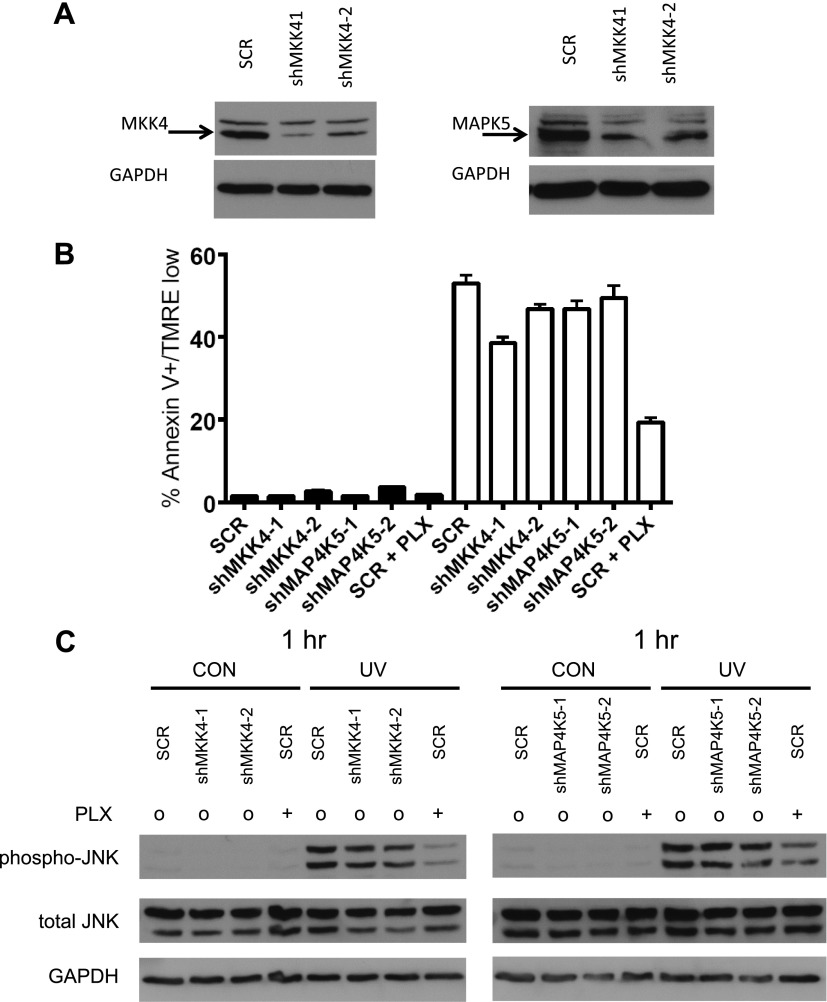
Figure 3—figure supplement 2. Single knockdown of MKK4 or MAP4K5 partially inhibits JNK activation and UV-induced apoptosis.

Figure 3—figure supplement 2.

(A) Western blot of HaCaT cells expressing two shRNA clones against MKK4 and MAP4K5 show significant knockdown of targets proteins (shMKK4-1: 89.3%, shMKK4-2: 71.9%, shMAP4K5-1: 86.4%, shMAP4K5-2: 84.1%; ImageJ). (B) HaCaT cells, expressing non-silencing scramble-shRNA (‘SCR’), shMKK4-1, shMKK4-2, shMAP4K5-1, or shMAP4K5-2 were either unirradiated (black bars) or irradiated (open bars) with 720 J/m2 of UVB in the absence (‘o’, 1:2000 DMSO) or presence (‘+’) of 1 μM PLX4720 and analyzed by FACS for apoptosis (Annexin V+, TMRE-) at 24 hr. UV-induced apoptosis is suppressed most substantially by MKK4 (up to 27.3%), but not substantially by MAP4K5 (up to 11.6%) in HaCaT cells. These results are consistent with the fact that ZAK can account for the majority of the effect of BRAFi-induced suppression of JNK signaling. Importantly, since MKK4 is important for JNK activation, and ZAK activates MKK4, the partial suppression of phospho-JNK activation and apoptosis is expected. (C) HaCaT cells, treated as above, were processed for Western blots at 1 hr following UV exposure to assess JNK activation. Significant activation of phospho-JNK is still observed at 1 hr post-irradiation in all cell lines, as compared to SCR cells treated with PLX4720.
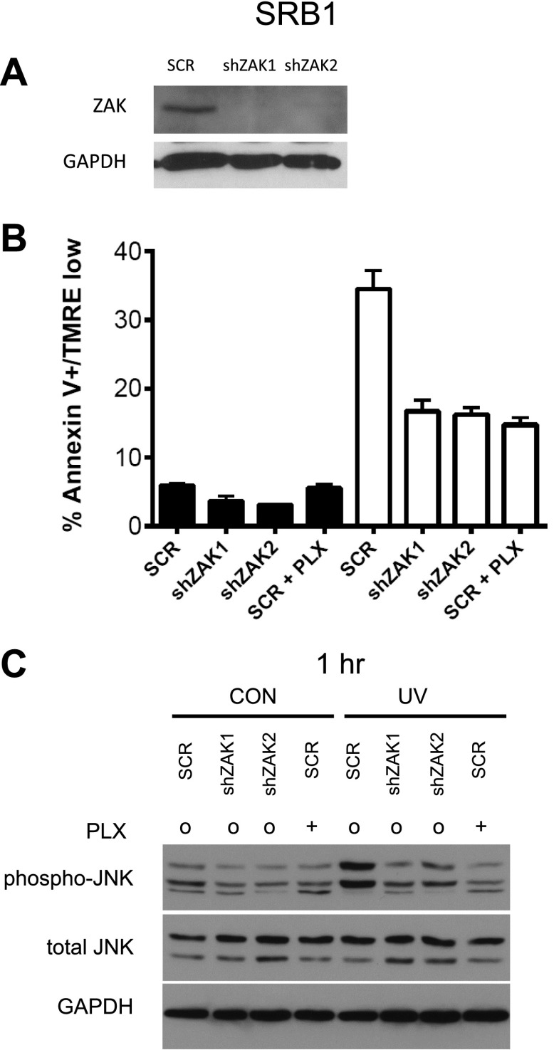
Figure 3—figure supplement 3. Knockdown of ZAK potently inhibits JNK activation and UV-induced apoptosis in SRB1 cells.

Figure 3—figure supplement 3.

(A) Western blot of SRB1 cells expressing two shRNA clones (shZAK1, shZAK2) all show significant knockdown of ZAK protein. (B) SRB1 cells, expressing non-silencing scramble-shRNA (‘SCR’), shZAK1, or shZAK2, were either unirradiated (black bars) or irradiated (open bars) with 720 J/m2 of UVB in the absence (‘o’, 1:2000 DMSO) or presence (‘+’) of 1 μM PLX4720 and analyzed by FACS for apoptosis (Annexin V+, TMRE-) at 24 hr. UV-induced apoptosis is significantly suppressed by both ZAK shRNA clones in SRB1 cells. shZAK1/2-expressing SRB1 cells and PLX4720-treated SRB1 scrambled-shRNA-expressing cells show similar degrees of suppression (90%, 92.5% of drug-treated cells), again consistent with the fact that ZAK can account for the majority of the effect of BRAFi-induced suppression of JNK-dependent apoptosis. (C) SRB1 cells, treated as above, were processed for Western blots at 1 hr following UV exposure to assess JNK activation. Significant suppression of phospho-JNK is observed at 1 hr post-irradiation in all cell lines where ZAK is knocked down, as well as in SCR cells treated with PLX4720.
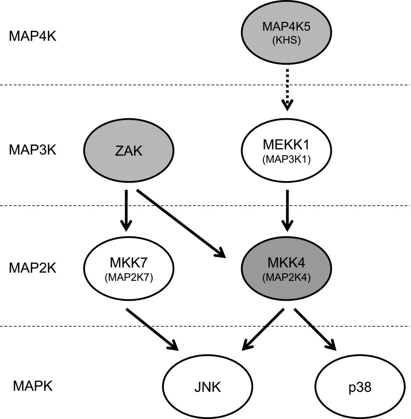
Figure 3—figure supplement 4. Vemurafenib and PLX4720 inhibit multiple kinases upstream of JNK and p38.

Figure 3—figure supplement 4.

The schematic shows MAP kinases upstream of JNK and p38 that are inhibited by these BRAF inhibitors (gray-shaded). Vemurafenib and PLX4720 inhibit ZAK (principally) and MKK4 (MEK4/MAP2K4), resulting in inhibition of MKK7 and MKK4 and, ultimately, JNK. p38 activation was diminished by drug exposure in some contexts , but not to the degree that JNK activation was. Vemurafenib and PLX4720 also inhibit MAP4K5, which has been shown to be upstream of MKK4 and JNK.