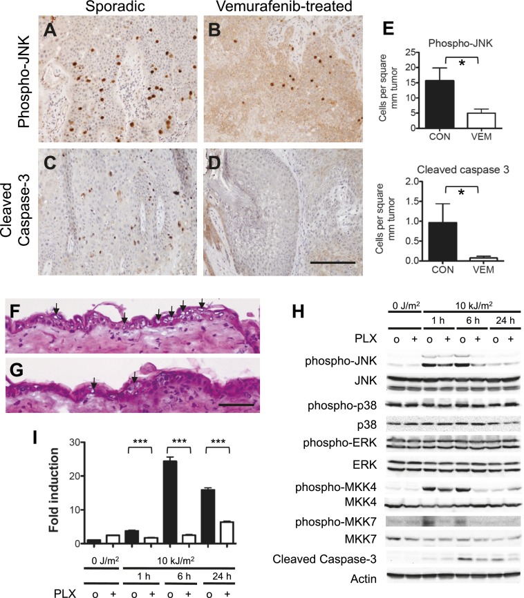
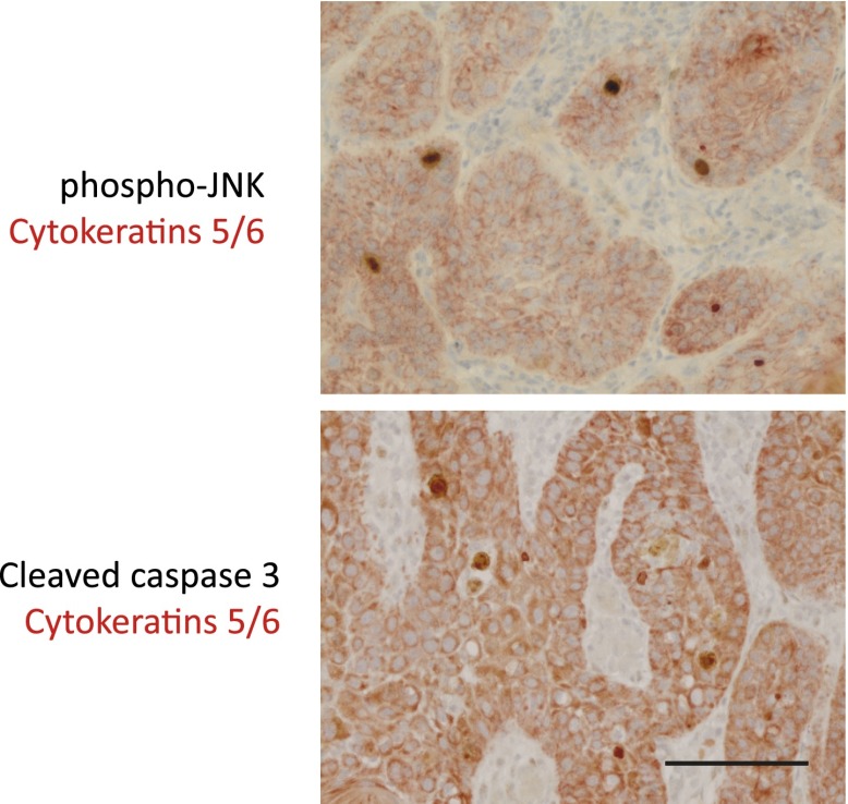
Figure 4. Vemurafenib and PLX4720 suppress apoptosis and JNK signaling in vivo.
(A–D) cSCC samples from vemurafenib-treated patients and non-treated patients were analyzed by immunohistochemistry for phospho-JNK and cleaved caspase-3 expression. cSCC arising in vemurafenib-treated patients show decreased expression of phospho-JNK (B) and cleaved caspase-3 (D) as compared to sporadic cSCC in patient never treated with vemurafenib (A and C). Scale bar is 100 μm. (E) Comparisons of stained cells normalized to mm2 of tumor area revealed significant suppression of both phospho-JNK and cleaved caspase 3 expression in vemurafenib-treated cSCC (‘*’, p<0.05). (F and G) Hematoxylin-stained cryosections of skin harvested at 24 hr post-irradiation showed extensive apoptosis (arrowheads) with vacuolated blebbed cells and clumped pyknotic nuclei in control-treated mice (F) and significantly fewer apoptotic cells in PLX4720-treated mice (G). Scale bar is 50 μm. (H–I) Vehicle-treated (‘o’) and PLX4720-treated (‘+’) mice were unirradiated or irradiated once, and epidermis was harvested at 1 hr, 6 hr, and 24 hr post-irradiation. (H) Significant UV-induced upregulation of both phospho-JNK and phospho-p38 were observed within 1 hr, with significant suppression of phospho-JNK in PLX4720-treated mice by 6 hr and minimal suppression of phospho-p38. Phospho-ERK levels remained constant. The upstream regulators of JNK, MKK4 and MKK7, were both significantly activated within 1 hr of irradiation, and potently suppressed in PLX4720-treated mice. Cleaved caspase-3 levels increased within 6 hr and were suppressed in PLX4720-treated mice. (I) Noxa was induced most significantly at 6 hr and was potently suppressed by PLX4720 at all time points (‘***’, p<0.001).


