
Figure 1.
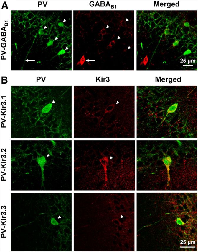
Immunoreactivity for GABAB1 and Kir3 subunits in hippocampal PVIs. A, Confocal images of the cell body layer of the hippocampal CA1 area showing immunofluorescent labeling for PV (left, green) and GABAB1 (middle, red) and their colocalization in the superimposed image (right). Note the strong immunolabeling for the GABAB1 subunit in the soma of a PV-negative interneuron (arrow) and the lower but discernible labeling in PV-positive somata (arrowheads). B, Confocal images illustrating the colocalization of PV (left, green) with Kir3.1 (top row, middle, red), Kir3.2 (middle row, middle), and Kir3.3 subunits (bottom row, middle). Immunoreactivity for all the three Kir3 subunits is present in PV-positive interneurons in and near the CA1 str. pyramidale (arrowheads).