Figure 2.
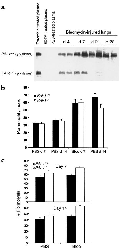
Dynamics of fibrin accumulation in bleomycin-exposed mice. Bleomycin (2.5 U/kg) or PBS was instilled intratracheally into PAI-1–/– and PAI-1+/+ mice. (a) To measure tissue fibrin content, lungs were harvested from heparinized mice on the days indicated and were processed as described in Methods. As positive and negative controls, fibrin samples formed within thrombin-treated plasma and anticoagulated plasma were prepared and processed in an identical manner. Samples were separated by SDS-PAGE and then immunoblotted to detect plasmin-generated, fibrin-derived γ-γ dimer fragment. (b) Vascular permeability was measured 7 days and 14 days after bleomycin administration, using lung accumulation of Evans blue dye that was injected intravenously 1 hour before sacrifice. Data are expressed as mean ± SEM; n = 7 for bleomycin-injured mice, n = 3 for PBS control mice. (c) The rate of fibrin degradation was measured in lungs of PAI-1–/– and PAI-1+/+ mice 7 days and 14 days after intratracheal instillation of PBS or bleomycin. At the time of sacrifice, fibrin was formed within pulmonary airspaces by intratracheally instilling fluorescein-labeled fibrinogen, plasminogen, and thrombin. After 5 hours, the percent of soluble fluorescent material was measured. Data are expressed as mean ± SEM; n = 4–7 mice per group.

