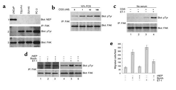
Figure 1.
NEP expression, FAK phosphorylation, and cell migration in PC cells. (a) Panel 1: total cell lysates (20 μg) from PC cells were analyzed for NEP protein by Western blot as described in Methods using the anti-NEP antibody 5B5. Panel 2: 300 μg of PC total cell lysates were immunoprecipitated (IP) with anti-FAK antibody C-20, separated by SDS-PAGE, transferred to nitrocellulose, and Western blotted with anti-pTyr mAb PY20. Panel 3: the same blot shown in panel 2 was stripped and reprobed with antibody C-20 for FAK protein. (b) LNCaP cells cultured in RPMI containing FCS and the NEP competitive enzyme inhibitor CGS24592 (CGS) for 2 hours at concentrations ranging from 0 to 100 nM were immunoprecipitated with anti-FAK antibody C20, separated by SDS-PAGE, transferred to nitrocellulose, and Western blotted consecutively with mAb PY20 (upper panel) and antibody C-20 (lower panel). (c) LNCaP cells cultured in RPMI without serum (lane 1), 10 nM ET-1 for 20 minutes (lane 2), 10 nM CGS24592 for 2 hours (lane 3), or 10 nM CGS24592 for 2 hours followed by 10 nM ET-1 for 20 minutes (lane 4). Cells were lysed and analyzed as described in b. (d) TSU-Pr1 cells were cultured in media without FCS for 24 hours (lanes 1 and 4), followed by the addition of 10 nM bombesin (Bomb., lane 2) or 10 nM ET-1 (lane 5) for 20 minutes; or by the addition of 50 μg/ml of rNEP for 2 hours, and then bombesin (lane 3) or 10 ET-1 (lane 6) for 20 minutes. Cells were lysed and analyzed as described in b. (e) Cell migration assays were performed in conditions identical to d. Bars represent SD. Experiments were repeated three times with similar results.

