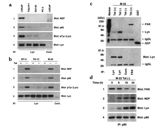
Figure 6.
Phosphorylated Lyn associates with NEP and p85. (a) 500 μg of total cell lysates were immunoprecipitated with rabbit anti-Lyn antibody or control rabbit antibody, separated by SDS-PAGE, transferred to nitrocellulose, and Western blotted with anti-NEP antibody, anti-p85 antibody, anti-pTyr antibody or mouse anti-Lyn antibody. (b) WT-5, TN-12, and M-22 cells were cultured with (+) and without (–) 1 μg tetracycline. Cells were lysed and analyzed as described in a. (c) Upper panel: M-22 cells were cultured with or without (–) 1 μg tetracycline for 48 hours. Cells were lysed and Far Western blotted as described in Methods. FAK immunoprecipitate of M-22 cells was used as a positive control for COOH-terminal SH2 domain of p85 to bind. GST; 0.1 μg recombinant GST protein used as a positive control for anti-GST antibody. Cont.; negative control by rabbit control antibody. IgGL; IgG light chain. (c) Lower panel: the same blot shown in upper panel was stripped and reprobed with mouse anti-Lyn antibody. (d) M-22 cells were cultured without (–) 1 μg tetracycline for various time periods. Cell lysates were immunoprecipitated with rabbit anti-p85 antibody, separated by SDS-PAGE, transferred to nitrocellulose, and Western blotted with anti-FAK antibody, anti-NEP antibody, mouse anti-Lyn antibody, or anti-p85 antibody.

