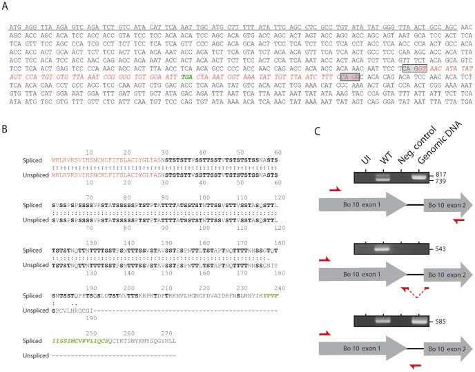
Figure 1. Bo10 mRNA undergoes alternative splicing.
A. Nucleotide sequence of BoHV-4 Bo10. The underlined nucleotides represent the sequence encoding the predicted signal peptide. The boxes indicate the splicing donor and acceptor sites. The sequence in red italics indicates the nucleotides removed after splicing. Nucleotides in green indicate the STOP codon in the intron. B. Amino acid comparisons of the predicted spliced and unspliced Bo10 products. The red amino acids represent the signal peptide sequence. The green amino acids indicate the predicted transmembrane region of the Bo10 spliced expression product. Serines and threonines are highlighted in bold. C. RT-PCR analysis of the BoHV-4 Bo10 reading frame. MDBK cells were mock infected or infected with the BoHV-4 V.test strain at a MOI of 1. Twenty-four hours p.i., expression of Bo10 was studied using different pairs of primers specific for the spliced and/or the unspliced Bo10 products. Red arrows indicate locations of the different primers. Uninfected cell samples (UI), reactions without reverse transcriptase (Neg. control) and amplification of viral genomic DNA are provided as controls. Sizes in bp are indicated on the right.

