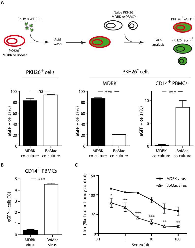
Figure 6. Myeloid virions are more infectious for CD14+ PBMCs but are more sensitive to antibody neutralization.
A. Infection by co-cultures. MDBK or BoMac cells previously stained with the membrane dye PKH26 were infected with BoHV-4 WT BAC virions (MOI of 1). Three hours p.i., cells were washed with acidic solution (PBS pH 3) in order to inactivate and remove the inoculum, and 12 hours p.i., freshly isolated PBMCs or MDBK controls were added. After 24 hours of co-cultivation, cells were collected and the proportion of eGFP expressing cells was measured in PKH26+ and PKH26− cells. For PBMC co-cultures, the proportion of eGFP expressing cells was measured in PKH26− CD14+ cells. The data presented are the average ± SEMs for 5 measurements and were analyzed by Student's t-test, *** p<0.001. B. BoHV-4 WT BAC cell-free virions propagated on MDBK or BoMac cells were added to bovine PBMCs at a MOI of 1 according to titers measured on MDBK cells. 24 hours later, percentages of eGFP positive cells were measured in CD14+ PBMCs by flow cytometry. The data presented are the average ± SEMs for 5 measurements and were analyzed by Student's t-test, *** p<0.001. C. BoHV-4 WT BAC virions propagated on MDBK or BoMac cells were incubated with sera of 3 different rabbits infected with BoHV-4 V.test strain (propagated on MDBK cells). After incubation (2 h, 37°C), the viruses were plaque assayed for infectivity on MDBK cells. BoHV-4 titers are expressed relative to virus without antibody. The data presented are the average ± SEMs for 3 measurements and were analyzed by 2way ANOVA and Bonferroni posttests, * p<0.05, ** p<0.01, *** p<0.001.

