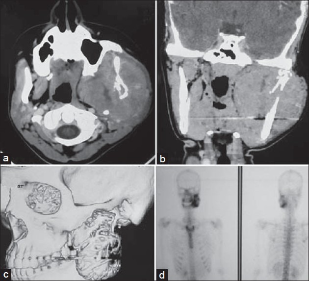
Figure 2.

CT scan: (a) Axial section, (b) Coronal section, and (c) Reconstructed image showing a heterodense mass lesion measuring 8 × 7 cm at its greatest dimension, causing bony destruction of the ascending mandibular ramus, with extension into the skull base. The tumor caused effacement of the fat and muscle planes of the infratemporal fossa and the masticator space, (d) A bone scintigram showed an increased abnormal activity in the left side of the mandible corresponding to the tumor
