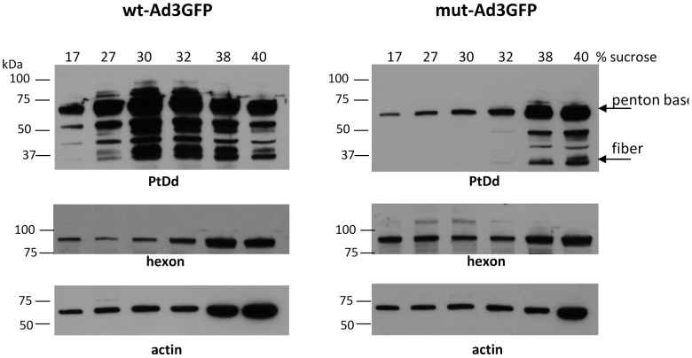
Figure 3. Detection of PtDd in infected cells.
HeLa cells were infected with wt-Ad3GFP or mu-Ad3GFP at an MOI of 500 VP/cell. Thirty six hours later cells were collected and cell lysates subjected to ultracentrifugation in a 15–40% sucrose step gradient. Fractions with different densities were then analyzed by Western blot using polyclonal antibodies raised against purified PtDd. The antibody recognizes Ad3 penton base (61.8 kDa) and fiber (34.8 kDa). The band at ∼50 kDa is most likely a fiber derivative. Blots were also hybridized with anti-Ad antibodies that recognize Ad3 hexon (∼90 kDa) and with anti-actin antibodies (loading control).

