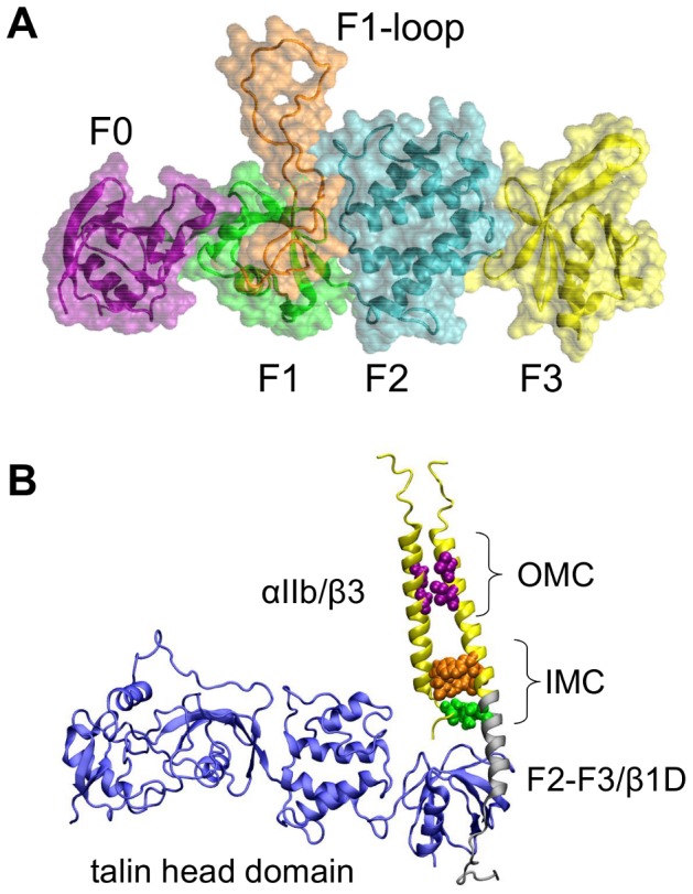
Figure 1. Talin and the talin/integrin TM complex.

A. Structure of talin (F0–F3; PDB:3IVF), showing the F0 domain in purple, the F1 domain in green, the F1 insertion in orange, the F2 domain in cyan, and the F3 domain in yellow. The loop inserted in the F1 domain was generated using Modeller. B. Model of the talin/αβ complex. The β chimeric peptide was comprised of β3 residues 688–719/β1D residues 753–787. The part of the structure corresponding to the αIIb/β3 structure (PBD:2K9J) is shown in yellow, the F2–F3/β1D (PDB:3G9W) is in grey, and the talin head domain (PDB:3IVF) is in blue.
