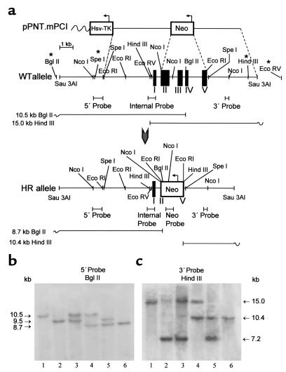
Figure 1.
Disruption of the PCI gene. (a) Genomic organization of the PCI allele in 129S/v mouse strain. Polymorphic restriction sites, present in the majority (more than 90%) of analyzed Swiss mice, are indicated by asterisks. Black boxes in the genomic structure represent exon sequences. Upon homologous recombination, the neo gene replaces a 3.6-kb genomic fragment consisting of a major part of exon II, exons III and IV, and a major part of exon V, leading to complete disruption of the PCI gene. WT, wild type; HR, homologously recombined. (b) Southern blot analysis of mouse genomic DNA digested with BglII and hybridized to a 5′ flanking probe. The appearance of a novel 8.7-kb band indicates correct targeting at the 5′ end of the gene. (c) DNA digested with HindIII and hybridized to a 3′ semi-internal probe C. The appearance of a novel 10.4-kb band indicates correct targeting at the 3′ end of the gene. In b and c, the presence of two fragments differing in size in wild-type mice reflects polymorphism of the PCI allele occurring in the Swiss mice population. Lane 1, PCI+/+ mice with nonpolymorphic wild-type PCI alleles; lane 2, PCI+/+ homozygous mice with polymorphic wild-type PCI alleles; lane 3, PCI+/+ mice with polymorphic and nonpolymorphic wild-type PCI allele; lane 4, PCI+/– mice with nonpolymorphic wild-type PCI allele; lane 5, PCI+/– mice with polymorphic wild-type PCI allele; lane 6, PCI–/– with targeted PCI alleles. Correct targeting of the clones was further checked with additional digests using internal and neo-specific probes (not shown), confirming correct targeting and excluding additional random integration of the targeting vector at other loci.

