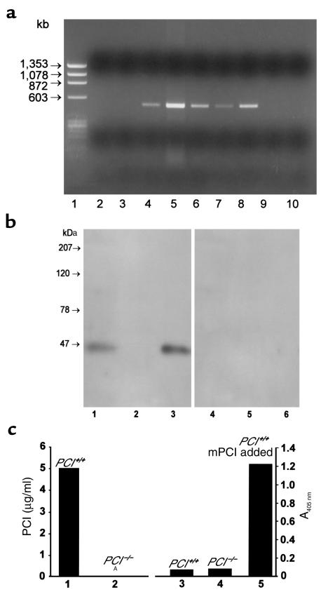
Figure 2.
(a) Tissue distribution of PCI mRNA determined by RT-PCR using 1 μg of RNA as template. Lane 1, PhiX 174/HaeIII marker; lane 2, testes of PCI–/– males; lane 3, ovaries of PCI–/– females; lane 4, seminal vesicles of PCI+/+ males; lane 5, testes of PCI+/+ males; lane 6, epididymides of PCI+/+ males; lane 7, prostates of PCI+/+ males; lane 8, ovaries of PCI+/+ females; lane 9, uteri of PCI+/+ females; lane 10, negative control. (b) Western blots. Crude Triton X-100 testis extracts (100 μg protein/lane) of PCI+/+ (lane 1) and PCI–/– (lane 2) mice are shown. Lane 3, recombinant mPCI (50 ng/lane). The samples in lanes 4–6 correspond to the samples in lanes 1–3, but they were incubated with preimmune serum only. (c) ELISA. PCI antigen concentration in crude Triton X-100 testis extracts (10 mg of total protein/ml) of PCI+/+ (lane 1) and of PCI–/– (lane 2) mice. ANo measurable antigen. Lane 3, A405 nm of 50% serum from PCI+/+ mice; lane 4, A405 nm of 50% serum from PCI–/– mice; lane 5, A405 nm of 50% serum from PCI+/+ mice with addition of 200 ng/ml recombinant mPCI (obtained value correlated with standard curve of the assay; not shown).

