Figure 4.
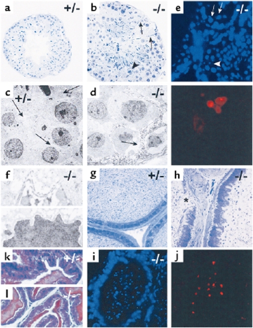
Histological analysis of male reproductive organs of PCI+/– and PCI–/– mice. (a–f) Testes of PCI+/– (a, c) and PCI–/– (b, d–f) mice are shown. (a, b) Semithin sections of seminiferous tubules. (c, d) Electron micrographs of seminiferous tubules. Whereas the seminiferous tubule of the PCI+/– mouse shows normal morphology (a), the seminiferous tubule of the PCI–/– mouse (b) has several unusual characteristics. The lumen is filled with immature spermatogenetic cells. The cytoplasm of Sertoli cells appears degenerated (arrows in b and d). Necrotic cells are found in the tubule wall (arrowhead in b) and in the tubule lumen. In c and d, arrows mark the cytoplasm of Sertoli cells. TUNEL assay (e) reveals spermatogenic cells that are positive for apoptosis (arrows), whereas Sertoli cells are devoid of such a signal (arrowhead). Electron micrograph of Sertoli cells of PCI–/– mice (f) shows disrupted Sertoli cell barrier of (upper panel). The nucleus lacks characteristic morphologic features of apoptosis (lower panel). (g–i) Cauda epididymis of PCI+/– (g) and of PCI–/– mice (h and i). In h, the lumen is filled with immature and malformed cells and with cell fragments. The lining cells appear irregular and are occasionally absent (asterisk). TUNEL assay (i) shows many apoptotic cells in the lumen but not in the lining epithelium. The seminal vesicles of both PCI+/– mice (k) and PCI–/– mice (l) appear normal.

