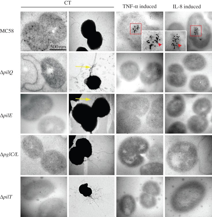
Figure 3.
Transmission electron micrographs showing uptake of human cytokines by Nm. TEM analyses of TNF-α- or IL-8-induced Nm wild-type strain and related mutants cultured for 9 h in the presence or the absence of 40 ng ml−1 TNF-α or IL-8. Untreated bacterial strains used as a negative control (CT). TNF-α-induced wild-type shows clear accumulation of gold particles inside the bacteria. The cells shown in this image are representative of approximately 72% of the analysed bacterial population (see electronic supplementary material, material and methods). IL-8-induced bacteria exhibit clear accumulation of intracellular gold particles. No uptake was observed in images of TNF-α- or IL-8-induced ΔpilE, ΔpilT or ΔpglC/L mutants, indicating that uptake of cytokines specifically require the glycosylated form of PilE or retracting pili. A small percentage (less than 0.1% of studied population) of the PilQ-deficient bacterial population could ingest the TNF-α or IL-8 in periplasmic space. Negative staining of ΔpglC/L glycosyl-transferase confirmed that Tfp formation in the mutant strain was equivalent to the wild-type strain. The yellow arrow shows the formation of an unknown form of pili by ΔpilE and ΔpilQ mutants, which may be functional to some degree. To confirm the specificity of anti-IL-8 or anti-TNF-α, the TNF-α-induced bacterial cells were detected with anti-IL-8 and vice versa, and with secondary antibody. No gold particles were detected in all examined strains and this was considered as an additional definitive negative control. Three independent experiments carried out in the presence and the absence of human recombinant cytokines are shown. The scale bar represents 500 nm and the insets represent 2.5 times magnification.

