FIGURE 3.

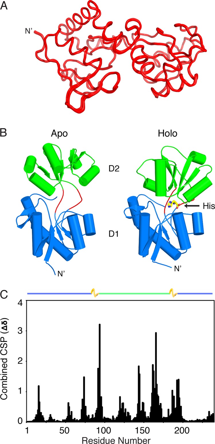
NMR structure of apo-HisJ. A, 30 lowest energy structures of apo-HisJ (red) overlaid (see Table 1 for structural statistics). B, side-by-side comparison of the lowest energy NMR structure of apo-HisJ and the crystal structure of holo-HisJ (PDB 1HSL). The first (D1) and second domain (D2) of each structure is colored blue and green, respectively. Holo-HisJ adopts a closed conformation in comparison with apo-HisJ, which results in histidine (yellow) being engulfed. Of note, hinge regions are colored red. C, combined NMR CSP for apo-HisJ and holo-HisJ (complexed with histidine). The domain organization of HisJ is noted above the figure where D1 subdomains (blue), D2 (green), and hinge regions (yellow) are indicated.
