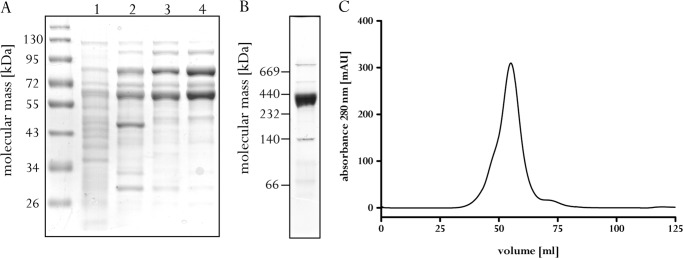
FIGURE 1.

Monitoring the purification and subunit composition of Acs/CODH from A. woodii. A, protein samples (10 μg) of each purification step were separated by SDS-PAGE and stained with Coomassie Brilliant Blue. Lane 1, cytoplasm; lane 2, sample after Q-Sepharose; lane 3, sample after phenyl-Sepharose; lane 4, sample after Sephacryl S-300. B, 15 μg of purified Acs/CODH was separated by clear native gel electrophoresis and stained with Coomassie Brilliant Blue. C, elution profile of Acs/CODH on Sephacryl S-300. mAU, milli-absorbance units.
