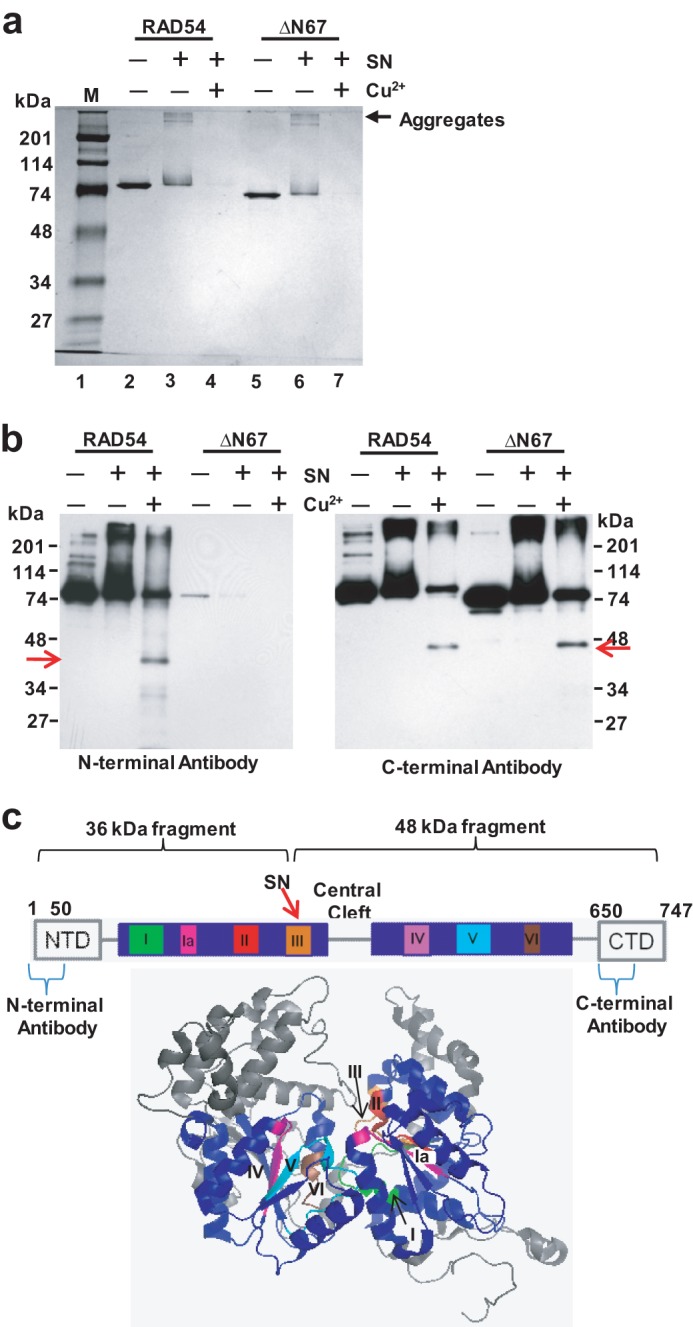
FIGURE 8.

Mapping of the fragments induced in RAD54 by SN in the presence of Cu2+. a, fragmentation and aggregation of 700 ng RAD54 and 700 ng RAD54 ΔN67 by 50 μm SN and 25 μm Cu2+. The reactions were carried out for 30 min at 37 °C, and the products were visualized by electrophoresis on a 10% SDS-polyacrylamide gel after staining with Coomassie Blue. M denotes Bio-Rad prestained SDS-PAGE standards broad range. b, specific RAD54 fragments (arrows) were identified by Western blotting using N-terminal- or C-terminal-specific RAD54 antibodies. c, schematic of RAD54 highlighting the seven conserved helicase motifs and the predicted binding site of SN. Shown below is the x-ray crystal structure of zebrafish RAD54 (PDB code 1Z3I). The seven conserved helicase motifs are highlighted in various colors in the two motor domains labeled in blue.
