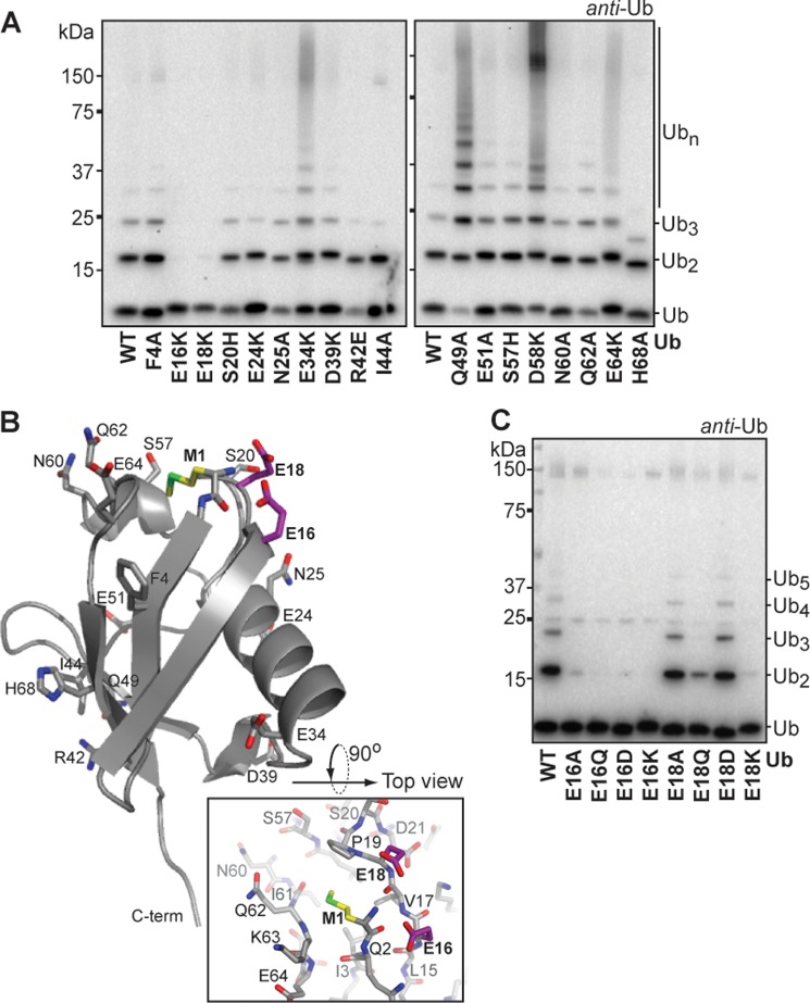
FIGURE 2.
Ubiquitin residues E16 and E18 are critical for ubiquitin chain formation. A, ubiquitin chain formation by HOIPRBR-LDD with different ubiquitin point-mutants in 1 h reactions. B, crystal structure of ubiquitin (PDB code 3PRM, chain d), illustrating the position of the tested ubiquitin surface point mutations in sticks (Met-1 (M1) in yellow, E16 and E18 in purple). The top view of the ubiquitin structure (PDB code 3PRM, chain d) illustrates the local environment of Met-1. C, HOIPRBR-LDD mediated chain formation with E16 and E18 ubiquitin point mutants. Reactions were stopped after 1 h.

