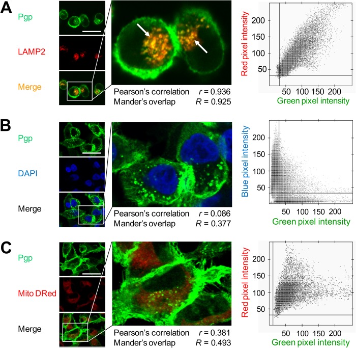
FIGURE 2.
Intracellular Pgp is localized to lysosomes in KBV1 (+Pgp) cells. A, Pgp colocalizes with lysosomal-associated membrane protein 2 (LAMP2, a well characterized lysosomal marker, arrows), whereas no colocalization was observed with DAPI (nuclear marker) (B) or MitoTracker® Deep Red (Mito Dred, mitochondrial marker) (C). The scatter plots, Pearson's correlation coefficient, and Mander's overlap coefficient in A–C were calculated using Imaris and Axiovision software from intracellular compartments of all cells. Photographs are typical from three experiments (A–C). Scale bars = 50 μm.

