FIGURE 6.
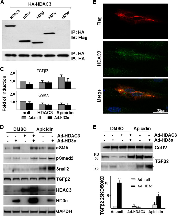
HD3α modulates HDAC3 function. A, Western blot analysis detected the association of HDAC3 isoforms with HDAC3 in HEK293 cells. All isoforms except for HD3δ associated with HDAC3. B, HD3α physically associated with and stabilized HDAC3. HAECs were infected with Ad-HD3α at an MOI of 10 for 6 h in complete growth medium and cultured in serum-free medium for 24 h, followed by immunofluorescence staining with anti-HDAC3 (green) and anti-FLAG (for HD3α; red). C–E, effect of overexpression or suppression of HDAC3 on HD3α-induced EndMT. HAECs were co-infected with Ad-HD3α (10 MOI) and Ad-HDAC3 (10 MOI) or in the presence of 1 × 10⁻⁵ mol/liter apicidin in complete growth medium for 6 h and then cultured in M199 medium supplemented with 5 ng/ml insulin for 24 h, followed by quantitative RT-PCR analysis of TGFβ2 and αSMA mRNA levels (C), Western blot analysis of cell lysates (D), and Western blot analysis of conditioned media with the ratio of cleaved to precursor TGFβ2 indicated in the bottom panel (E). Ad-null is included as a control and to normalize variations in MOI. DMSO was included as a vehicle control. *, p < 0.05; **, p < 0.01. Data presented are representative of or the average of three independent experiments. IB, immunoblot; IP, immunoprecipitation.

