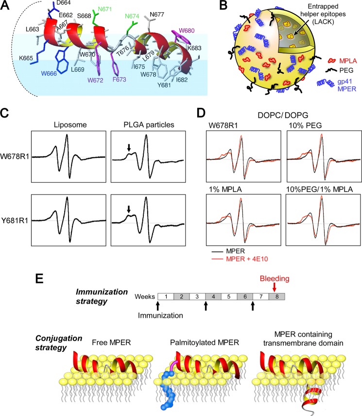
FIGURE 1.
Structural configuration of MPER segments in liposome vaccines. A, NMR structure of the HxB2 MPER in a virion mimic membrane surface. Residues essential for BNAb neutralization are color-coded as follows: blue for 2F5, green for Z13e1, and magenta for 4E10. B, schematic of a standard liposome vaccine formulation, including the various components used in immunization studies. C, comparison of EPR spectra of spin-labeled MPER Trp-678(R1) and Tyr-681(R1) peptides on liposomes versus poly(lactide-co-glycolide) (PLGA) particles. Arrows in the latter highlight immobilization. D, EPR spectra of a spin-labeled MPER Trp-678(R1) peptide bound to liposomes with varied PEG and MPLA compositions. Spectra were obtained in the absence (black) and presence (red) of 4E10. E, immunization schedule and various conjugation strategies employed for MPER display on the surface of the liposome vaccines. For clarity, only the outer leaflet of the membrane is shown.

