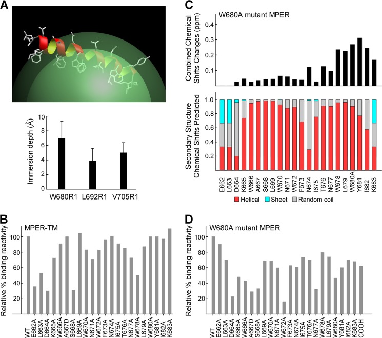
FIGURE 6.
Shifted immunogenicity of MPER-TM and W680A mutant MPER segments as a result of the loss of the accessible Trp-680 antibody-binding “hot spot.” A, MPER segment of the MPER-TM peptide is shown in ribbon representation on a micelle surface (top) to illustrate NMR secondary structural analysis with the corresponding EPR membrane immersion depth measurements of spin-labeled residues (bottom). B, representative epitope specificity of anti-MPER-TM polyclonal IgG antibody shown as binding of alanine mutants relative to the wild type sequence. C, combined 15N/1H amide chemical shift changes of the W680A mutant MPER, indicating local perturbations near the C terminus (top) with similar overall secondary structures (bottom) relative to the wild type MPER peptide. D, epitope mapping of W680A mutant MPER specific antibody. The purified polyclonal IgG antibody was tested for binding affinity by Biacore. Data are representative of three independent immunizations of a group of five mice each with W680A mutant MPER/liposomes.

