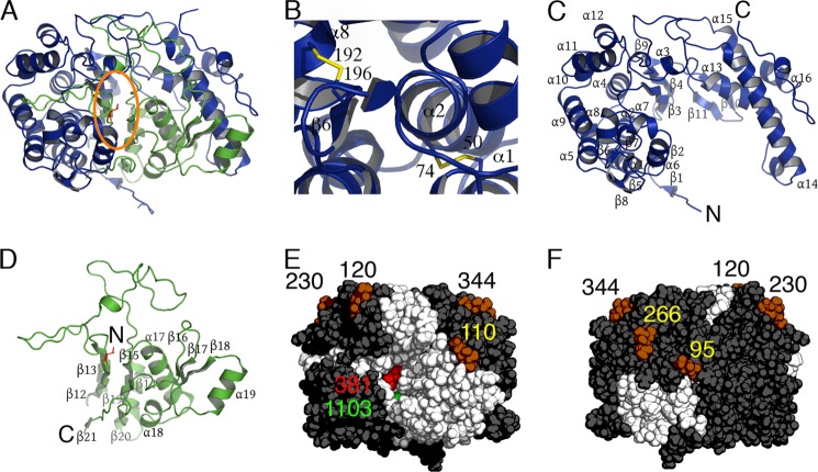
FIGURE 3.
Crystal structure of human GGT1. A, ribbon representation of the hGGT1 heterodimer. The large subunit (chain A) is colored blue, and the small subunit (chain B) is colored green. The active site Thr-381 is colored red. The orange oval outlines the active site cleft. B, view of the two disulfide bonds (Cys-50/Cys-74, Cys-192/Cys-196) in the large subunit of hGGT1. Ribbon representations of large (C) and small (D) subunits of hGGT1 are shown. E, the van der Waals surface of hGGT1 with the active site cleft facing the viewer. Thr-381 is colored red. The large subunit is colored dark gray, and the small subunit is white. The six GlcNAcs on the surface are represented as dark orange van der Waals spheres and are labeled by the residue number of the asparagine to which they are anchored (black and yellow numbers). A green sphere represents the anion-binding site. F, backside of view in E.

