
FIGURE 2.
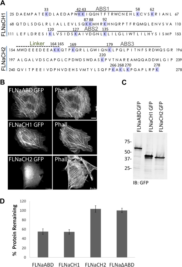
CH1 is the minimal fragment of FLNa sufficient for ASB2α-mediated degradation. A, primary amino acid sequence of FLNaABD. The ABD is composed of two calponin homology domains (CH1 and CH2) connected by a linker region. The predicted actin-binding sites (ABS1, ABS2, and ABS3) are indicated. Lysines are highlighted in blue, and the corresponding residue numbers are indicated above. B, CHO cells transfected with FLNaABD GFP, FLNaCH1 GFP, and FLNaCH2 GFP were fixed and stained for phalloidin (Phall). Scale bar, 10 μm. C, CHO cells transfected with FLNaABD GFP, FLNaCH1 GFP, and FLNaCH2 GFP were lysed and immunoblotted (IB) using anti-GFP antibody. D, CHO cells transiently expressing FLNaABD GFP, FLNaCH1 GFP, FLNaCH2 GFP, and FLNaΔABD GFP were transfected with either dsRed-ASB2α or dsRed-ASB2αΔS. 48 h after transfection, cells were detached and washed with PBS, and the GFP intensity of dsRed-expressing cells was assessed by flow cytometry. Bar chart depicts mean percentage of GFP-tagged protein remaining ± S.E. in dsRed-ASB2α-expressing cells normalized to levels in dsRed-ASB2αΔS-expressing cells (see “Experimental Procedures” for details). Data are from at least seven independent experiments.