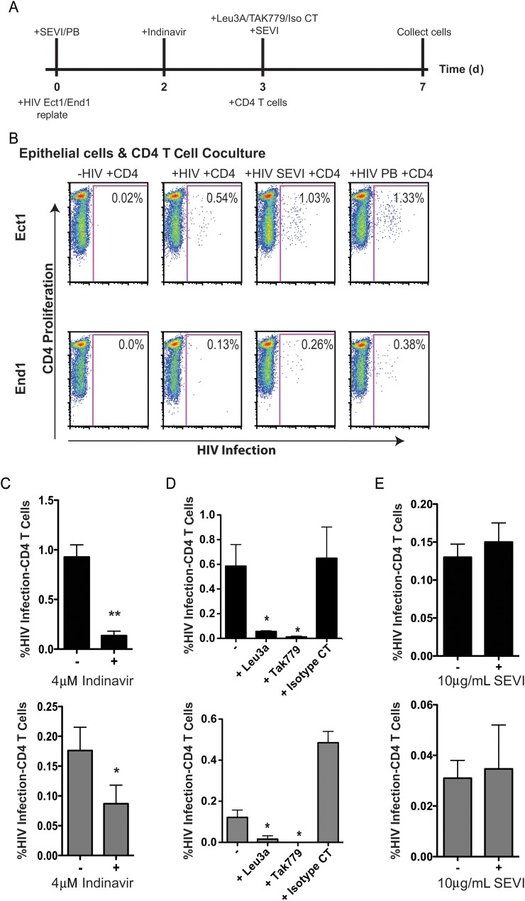
Figure 5.
Infected epithelial cells are capable of transmitting the infection to cocultured target CD4+ cells. Infected Ect1 and End1 cells (approximately 300 000 cells/well, 12-well plate) were cocultured with approximately 2 million activated CD4+ T cells per well in Roswell Park Memorial Institute complete media following coculture assay. A, Outline of experimental design that indicate when inhibitors or enhancing factors were added. B, CD4+ T-cell human immunodeficiency virus type 1 (HIV-1) infection (x-axis) vs CD4+ T-cell proliferation (y-axis). Infection of CD4+ T cells was enhanced when epithelial cells were incubated with HIV and semen-derived enhancer of virus (SEVI) fibrils at day 0 (Ect1, P = .041; End1, P = .02), or polybrene (PB; Ect1, P = .1; End1, P = .3) based on 3 separate experiments. Noninfected epithelial cells cocultured with CD4+ T cells acted as a negative control. C, Infection of CD4+ T cells was sensitive to inhibition by the viral protease inhibitor indinavir, indicating that new viral maturation was required to transmit the infection to CD4+ T cells. Ect1 (top, black graph) or End1 (bottom, gray graph) were treated with indinavir on day 2 prior to and during coculture (Ect1, P = .0074; End1, P = .005). D, Coculture infection of CD4+ T cells was inhibited by Leu3A (Ect1, P = .03; End1, P = .04) and TAK779 (Ect1, P = .03; End1, P = .04), indicating a CD4- and coreceptor-dependent infection. Inhibitors were added on day 3 prior to addition of CD4+ T cells. E, Addition of SEVI fibrils on day 3 prior to coculture with CD4+ T cells had no effect on coculture infection of CD4+ T cells. P values were determined using an unpaired, 2-tailed T test comparing infected epithelial cell coculture with inhibitor treated–infected coculture. Graphs show mean and standard deviation of 3 separate experiments. (*, **, *** indicate increasing degree of significance).

