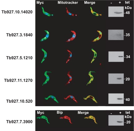
Supplementary Figure S1. Immunofluorescence analysis of the putative glycosomal membrane proteins in procyclic T. brucei.
N-terminally or C-terminally myc-tagged versions of the proteins were detected (green) by using a commercial Myc antibody (Sigma-Aldrich). Mitochondria were visualized (red) using mitotracker. The endoplasmatic reticulum (ER) was detected using an antibody directed against the ER lumen protein BiP (red). Overlays (Merge) of the green staining and the red staining are shown to visualize the common compartmentalization of the proteins. On the right side, western blots are shown to illustrate the expression of the myc-tagged membrane proteins. (+) and (-) indicate tetracycline-induced and -uninduced cells respectively.

