
FIGURE 2.
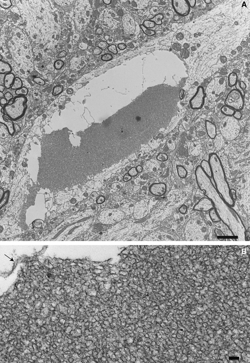
Thy1.2-hNIPA1G106R rat morphology. (A) A neurite within the anterior horn of the lumbar cord of a 12-week-old Thy1.2-hNIPA1G106R rat showing a partially digested large aggregate of a tubulovesicular structure within an autophagic vacuole. Scale bar = 2 μm. (B) Higher magnification shows the elongated tubular morphology of these aggregates. Arrow indicates the remnants of the surrounding double membrane. Scale bar = 100 nm.