
FIGURE 6.
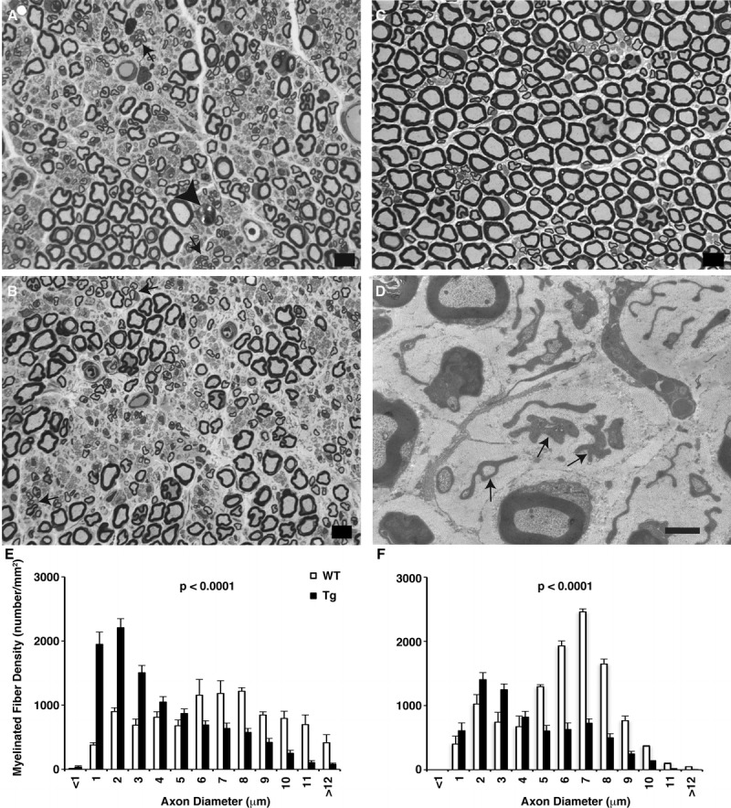
Thy1.2-hNIPA1G106R transgenic (Tg) rat peripheral nerve. (A) One-micrometer-thick toluidine blue–stained plastic sections from the proximal sciatic nerve of a Tg rat showing large myelinated fiber loss. Arrows point to clusters of thinly myelinated regenerating axonal sprouts; a fiber undergoing Wallerian degeneration is marked with an arrowhead. (B) Fiber loss was more prominent distally in the tibial nerve from a Tg rat. (C) Wild-type (WT) proximal sciatic nerve. Scale bar = 10 μm. (D) At the ultrastructural level, there were empty stacks of Schwann cell processes (arrows), some engulfing collagen, because of unmyelinated axon loss in the Tg nerve. Scale bar = 2 μm. (E, F) Myelinated fiber size distribution histograms of sciatic (E) and tibial (F) nerves from WT and Tg rats at 40 weeks of age. The composite histograms were derived from 3 rats in each group. Loss of large myelinated fibers (i.e. myelinated fibers with axon diameter >5 μm) in the Tg was more prominent distally in the tibial branch (F) compared with the proximal sciatic nerve (E). The prominent increase in the small myelinated fiber population seen at both levels reflects ongoing regeneration.