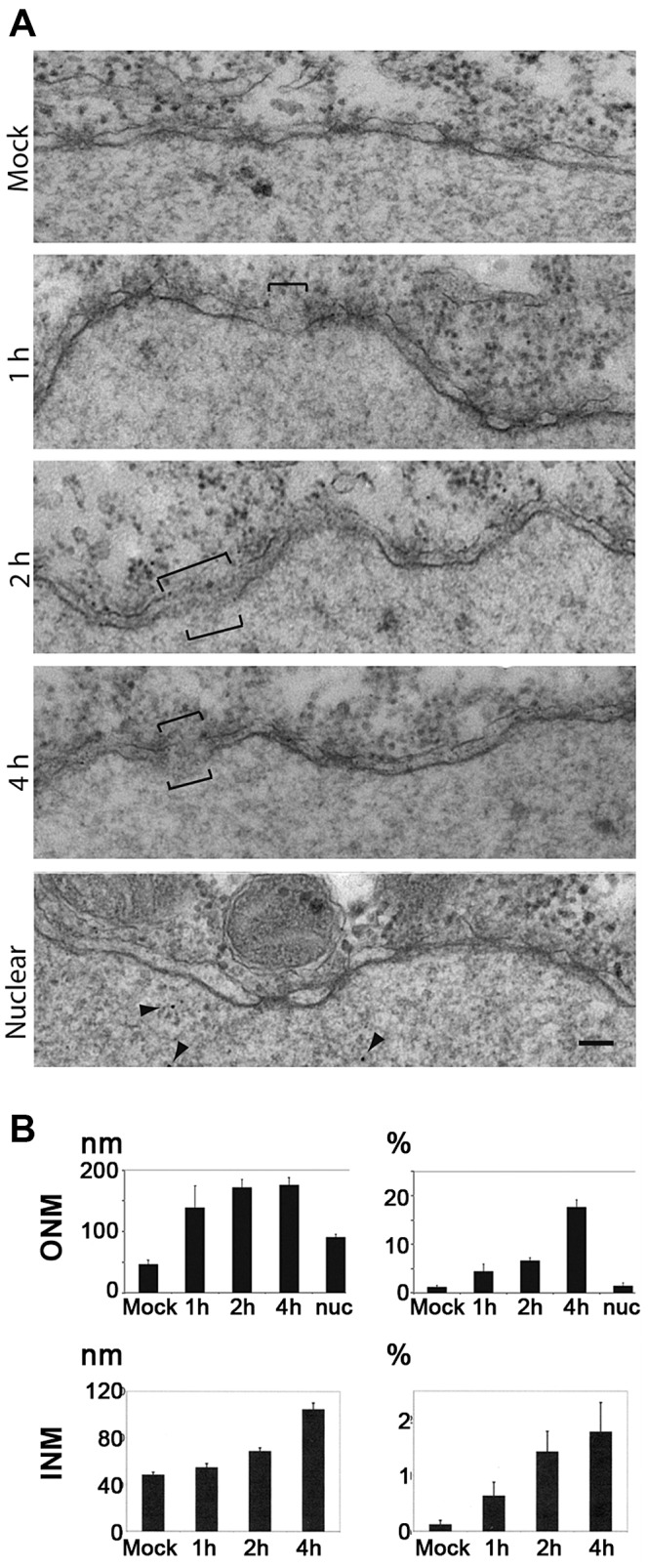
Figure 7. H1 causes nuclear envelope breaks in Xenopus laevis oocytes after microinjection into the cytoplasm.

A. Electron microscopy of the nuclear membrane after microinjection of 2.13×10⁵ pfu./ml, or 2.17×10⁸ genomes/ml H1 into the cytoplasm of Xenopus laevis oocytes. The oocytes were fixed after 1, 2, 4 h prior to preparation of the nuclei and staining. Membrane breaks are indicated by brackets, the nucleus is on the bottom of each panel. MOCK: Tris EDTA, pH 7.8 injection and incubation for 4 h. Middle panels: injection of H1 with the indicated incubation time. Nuclear: nuclear microinjection (4 h RT). Bar = 100 nm. B. Quantification from 30 electron micrographs derived from microinjection into three oocytes per condition. Mock: mock-injected control. Nuc: nuclear microinjection after 4 h. Left panels: the length of the breaks. Right panels: proportion of degraded nuclear envelope. Nuc: nuclear microinjection. In summary these data show that PV-mediated NEBD leads to disruption of inner and outer nuclear membrane in Xenopus laevis oocytes, supporting that PV-mediated NEBD is a evolutionary well conserved process. The breaks in the membrane indicate that Ca++ leaks out of the lumen between the two membranes. The observation that ONM breaks occur with higher frequency than observed for the INM implies that membrane disintegration starts at the ONM.
