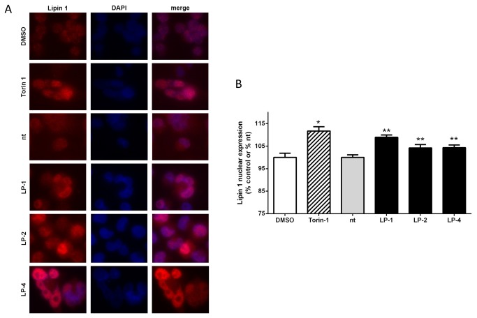
Figure 7. LPAAT-β siRNA treatment results in enhanced nuclear localization of Lipin 1.
Immunofluoresence staining (A) for Lipin 1 (red) with the nuclei counterstained with DAPI (blue) and overlay (merge) of MiaPaCa2 cells treated with: DMSO and 500 nM Torin-1 for 24 hours; control siRNA (nt), 10 nM LP-1, 25 nM LP-2 and 10 nM LP-4 for 48 hr. B) Quantitation of nuclear Lipin 1 fluoresence intensity from (A). Statistical significance was calculated using Student’s t-test: DMSO (*) vs Torin-1, p < 0.0001; siRNA nt (**) vs LP-1, p = 0.0005; vs LP-2, p = 0.05; vs. LP-4, p = 0.02. Results are representative of three independent experiments.

