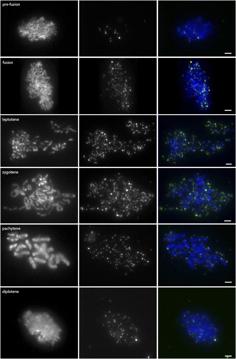
Figure 6.
Anti-gamma-H2AX localization on wild-type meiotic chromosome spreads. Images in the left column are DAPI-stained chromosome spreads; the stage of prophase is indicated on the image. Images in the middle column are of anti-gamma-H2AX counterstained with a TRITC-conjugated secondary antibody, and images in the right column are the color-combined images. Prefusion, n = 42; leptotene, n = 47; zygotene, n = 50; pachytene, n = 48; diplotene, n = 60. Scale bars represent 2 µm.

