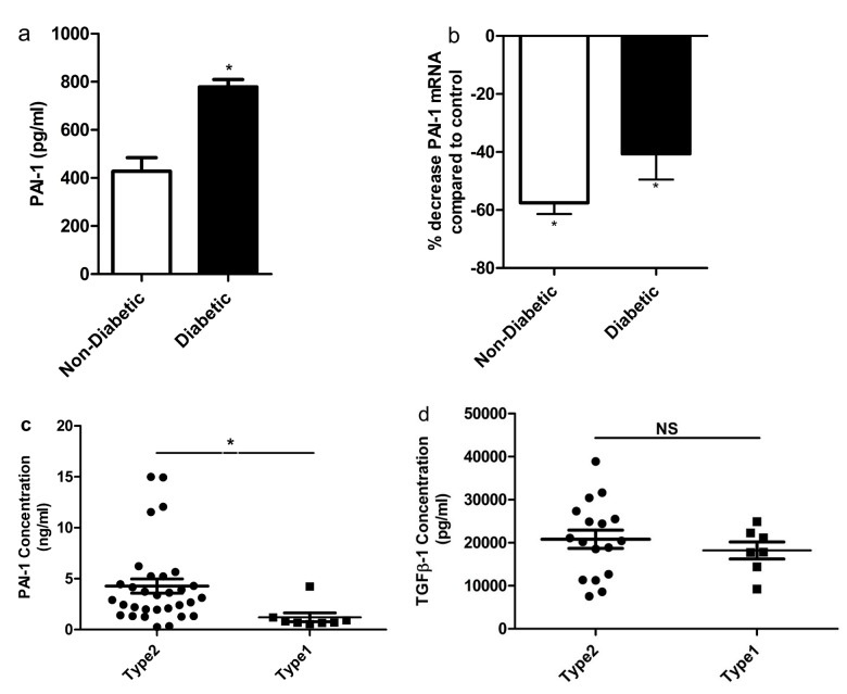
Figure 2. TGF-β1 mediates its action through PAI-1 in both diabetic and non-diabetic CD34+ cells.
(a) PAI-1 concentration in the conditioned media of diabetic and non-diabetic CD34+ cells. A significant increase in the secreted level of PAI-1 was observed in the conditioned media obtained from diabetic CD34+ cells compared to non-diabetic (p<0.05) (mean ± SEM; n=3). (b) Effect of control PMO and TGF-β1PMO on PAI-1 gene expression in CD34+ cells was assessed. Both diabetic and non-diabetic CD34+ cells were pretreated overnight with either scrambled PMO or TGF-β1PMO (40ng/ml). PAI-1 mRNA transcripts were quantified by real-time RT-PCR and was normalized to β-actin levels. Values in cells treated with scrambled PMO were set at 1.0. p<0.001(for non-diabetic compared to scrambled PMO treated cells); p=0.05 (for diabetic compared to scrambled PMO treated cells); n=10 for diabetic and n=3 for control. (c-d) Plasma concentrations of PAI-1 and TGF-β1 in type1 and type2 diabetic individuals. A significant increase (p<0.05) in the plasma concentration of PAI-1 in type2 diabetic individuals compared to type 1 (n=31 for type 2; n=7 for type 1), although the concentration of TGF-β1 was similar in both groups (20815.1pg/ml in type2 and 18212.2 pg/ml in type1). Each dot represents one patient sample.

