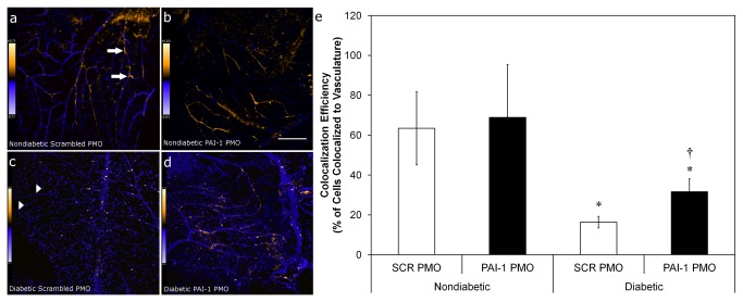
Figure 7. PAI-1 inhibition with PMO increases diabetic CD34+ cell homing to and association with vasculature in an acute I/R model.
CD34+ cells isolated from diabetic (n=8) or age- and sex-matched normal (n=4) donors were exposed to either scrambled (SCR) or PAI-1-specific PMO for 16hr prior to intravitreous injection 7 days post ischemic insult. Eyes were harvested 2 days later and processed for immunofluorescence and data was collected using laser scanning confocal microscopy. Panels (a-d) are typical fields that show calculated co-localization of CD34+ cells with retinal vasculature using Intensity Correlation Analysis (Mander’s coefiicient) and given false color to indicate the degree of co-localization probability (warmer colors = higher probability). Scale bar in (b) also applies to (a) and is 100 mm. Scale bar in (d) also applies to (c) and is 150 mm. (e) represents co-localization efficiency. Arrows indicate exogenously added cells that have not co-localized to vasculature (best seen in Panel C), while arrowheads (Panel A) indicate areas where exogenously added cells co-localize strongly (warm colors) with vessels. * = p<0.05 vs. non-diabetic cells with the same pre-treatment. † = p<0.05 versus SCR PMO with diabetic cells.

