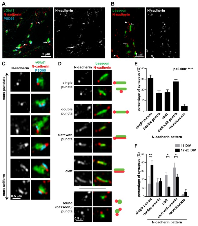
Figure 2. N-cadherin has a spectrum of localization patterns at the synaptic cleft.
(A) N-cadherin is localized between the pre- and post-synaptic compartments, represented by vGlut1 and PSD95 respectively. (B) N-cadherin is localized at or adjacent to the active zone, represented by bassoon. Arrows, N-cadherin puncta associated with synapses; arrowheads, N-cadherin puncta not associated with synapses. (C) N-cadherin localization at synapses varies from punctate, often flanking one side of the synapse, to uniform along the synaptic cleft. (D) Classification of N-cadherin localization patterns relative to bassoon into five categories (single puncta, double puncta, cleft with puncta, cleft, and round(bassoon)/puncta). Representative images and schematics of the five different N-cadherin localization patterns. (E) Percentage of synapses (mean±s.e.m) in 17-20 DIV hippocampal neurons in each N-cadherin pattern category. n=7 experiments, ≥77 synapses per experiment, 650 synapses total. p<0.0001, one-way ANOVA, Tukey’s post-test (p<0.01 for single puncta vs. double puncta; p<0.01 for single puncta vs. cleft; p>0.05 for single puncta vs. cleft with puncta; p<0.001 for single puncta vs. round/puncta). (F) Percentage of synapses (mean±s.e.m) in each N-cadherin pattern category in 11 DIV hippocampal neurons compared to matched cultures at 17-20 DIV. N-cadherin at synapses from neurons at 11 DIV is distributed more evenly along the synaptic cleft and less as puncta compared to synapses from neurons at 17-20 DIV. Two-way ANOVA with matched values and Bonferroni post-test. n=2 experiments, ≥103 synapses per experiment.

