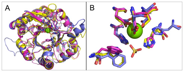
Figure 4. Structural alignment of SMases D.

L. laeta SMase D (1xx1) (purple) and the models for the SMase D-like proteins from the fungus A. flavus (yellow) and from C. pseudotuberculosis (blue), indicating the overall alignment (A) and the active site residues superposed in sticks (B), the Mg+2 ion as a green sphere and the co-crystalized SO4- ion in sticks.
