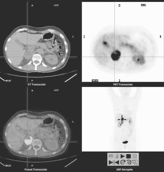
Fig. 3.

Right adrenal lesion. From the upper left, clockwise: computed tomography (CT), PET, MIP, and fused PET/CT images of l-6-[18F]fluoro-3,4-dihydroxyphenylalanine (18F-DOPA) PET/CT of a 51-year-old woman presenting with very high urinary normetanephrin levels. 123I-MIBG scintigraphy revealed only a mild to moderate uptake of the tracer in the right adrenal. Instead, 18F-DOPA PET/CT shows a very intense uptake in the right adrenal, consistent with pheochromocytoma. Note the concomitant uptake in the body–tail of the pancreas, and the intense uptake in the gallbladder. Images with no carbidopa premedication.
