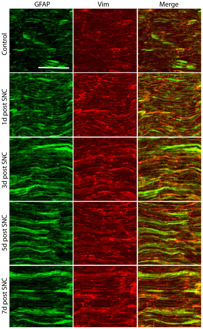
Figure 2. Expression of GFAP and vimentin in the lesioned sciatic nerve.
Low levels of GFAP and vimentin (Vim) were detected in the uninjured nerve. GFAP and vimentin are mainly expressed by Schwann cells. An increase in both GFAP and vimentin IR is apparent from 1 day after lesion. The picture was taken as a maximal projection and settings were adjusted to optimal levels in a similar fashion for all pictures taken. Scale bar, 50 µm.

