
Figure 2.
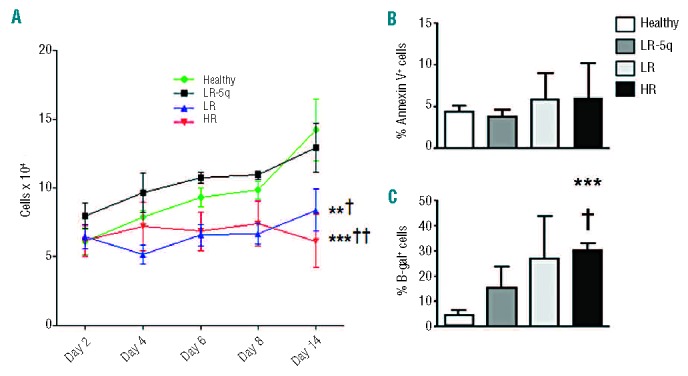
Proliferation of MDS-MSC is impaired. (A) Proliferation curves for each group were determined by seeding 5×10⁴ cells and measuring cell number at days 2, 4, 6, 8, and 14. (B) Apoptosis of MSC after 14 days of culture was determined by annexin V staining. (C) Senescence of MSC was demonstrated by β-galactosidase (β-gal) staining after 48 h of culture of 1×10³ cells per donor. Results are expressed as mean ± SEM of independent cases. For (A) numbers were Healthy=6, LR-5q=6, LR=6 and HR=5; for (B) Healthy n=6, LR-5q n=9, LR n=3 and HR n=3; (C) all groups n=4. Significance was set as */P≤0.05; **/††/‡‡P≤0.005; ***P≤0.001.