
Figure 6.
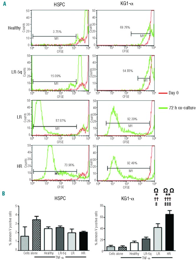
MSC from MDS patients induce different apoptotic and proliferative responses on healthy HSPC and KG1-α leukemic cells. (A) Proliferation of CD34+ healthy cells and KG1-α cells was determined using CFSE staining and co-culture with stroma with or without addition of 100 nM of TNF-α measuring the dilution of the dye after 72 h by flow cytometry. The histograms show the percentage of cells that diluted CFSE beyond the level at day 0 of representative examples in the non-adherent fraction after co-culture with TNF-α (results were similar in all fractions regardless of cytokine presence). (B) Effect on apoptosis after co-culture with stroma was only evident in KG1-α cells and in the fraction of cells that after 24 h were not attached to the MSC layers; the percentage of annex-in+ cells is shown for both HSPC and KG1-α in the non-adherent fraction (apoptosis was similarly low to that of cells without co-culture in all fractions with HSPC and in the adherent fraction of KG1-α). Ω indicates differences between cells alone with TNF-α and cells co-cultured with stroma. Results are expressed as mean ± SEM of independent cases. For (A) numbers were Healthy=6, LR-5q=6, LR=5 and HR=5; for (B)n=3 for HSPC experiments and n=4 for KG1-α for all groups. Significance was set as */‡/ΩP≤0.05; **/††/ΩΩP≤0.005; †††/‡‡‡P≤0.001.