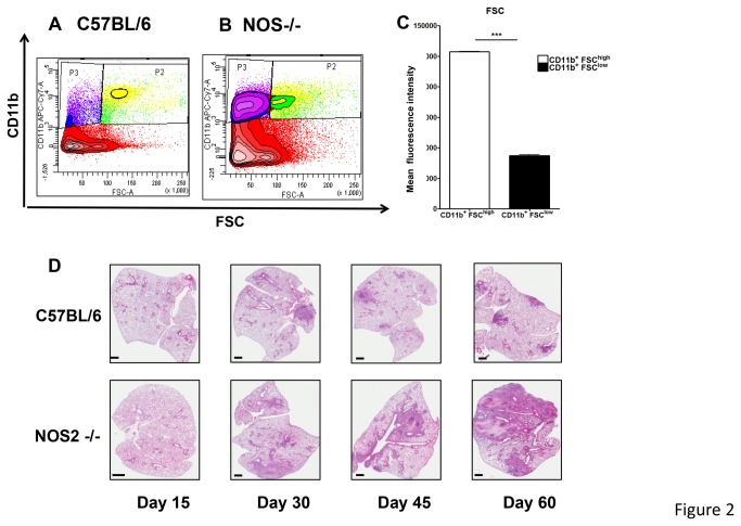
Figure 2. Two populations of CD11b+ cells based on FSC analysis.
As suggested by FSC analysis, the increased number of CD11b+ cells present in NOS2 -/- mice could be separated in two populations based on cell size (A, B). Both populations of CD11b+ FSChigh cells and CD11b+FSClow cells are significantly increased in NOS2 -/- mice compared to C57/BL/6 mice (A, B). Contour plot is representative of day 45 after infection. FSC analysis clearly identified two CD11b+ populations in the lungs of NOS2 -/- mice (C). Results are expressed as the mean values fluorescence intensity (± SEM, n=5) in the Lung, Student t-test, *** p<0.001. Necrotic granulomas are only present in the lungs of NOS2 -/- mice (D). At different time points after infection, lungs were harvested and stained with H&E after fixation and paraffin embedding. For both murine strains, lesions are not present at day 15 after infection. Starting day 30, necrotic granulomas become evident in the lungs of NOS2 -/- mice but not WT C57BL/6. As observed at day 45 and 60, necrotic granulomas coalesce leading to severe lung consolidation at day 60 when most NOS2 -/- are moribund.

