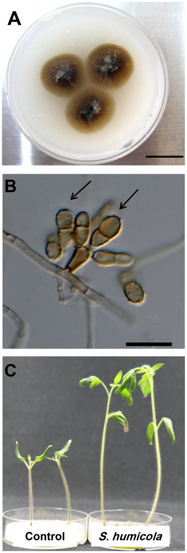
Figure 2. Scolecobasidium humicola colony and conidia.

Colony (A) and a light micrograph (B) of Scolecobasidium humicola after 14 days at 23°C grown on OMA medium. Arrowhead indicates a micronematous conidiophore with rough-walled septate conidia (arrows). Bars: A = 14 mm, B = 20 µm. (C) Tomato seedlings grown on basal media (OMA) amended with L-Leucine amino acid, the control on the right , and inoculated tomato seedlings with S.humicola on the left.
