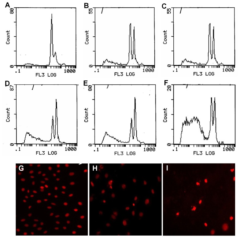
Figure 5. Revivification of RhoB gene and apoptosis of ovary carcinoma cells.
Flow cytometric analysis (FCM) together with fluorescence microscopy was adopted to assess apoptosis of ovary carcinoma cells after treated with TSA. Top and middle panels: when cells were treated for 10 h with 0 µmol/L (control), 0.05 µmol/L, 0.1 µmol/L, 0.25 µmol/L, 0.5 µmol/L and 1.0 µmol/L of TSA, the apoptosis rate revealed by FCM was 9.5% (A), 26.9% (B), 28% (C), 41% (D), 45.9% (E) and 66.9% (F) respectively. Bottom panel: PI stained fluorescence photomicrographs of ovary carcinoma cells after treated with TSA. Cells were treated for 10h with 0 µmol/L (G, control), 0.1 µmol/L and 0.5 µmol/L of TSA respectively, both 0.1 µmol/L (H) and 0.5µmol/L (I) of TSA could result in cellular morphological changes characterized as apoptosis: a brightly red-fluorescent condensed nuclei (intact or fragmented), reduction of cell volume, and apoptotic bodies.

