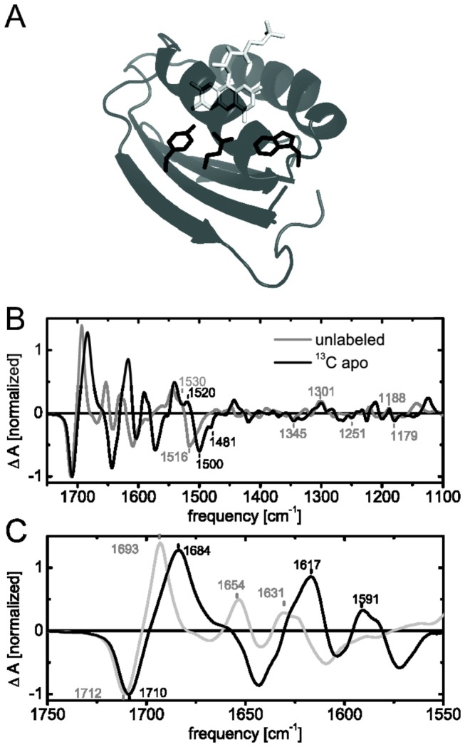
Figure 6. Selective unlabeling of the flavin chromophore (A, light grey) upon uniform 13C labeling of the protein (dark grey and black) using the riboflavin auxotrophic strain CpXribF.

Light-minus-dark FTIR difference spectra of unlabeled Slr1694 (grey) and apoprotein 13C-labeled Slr1694 (black) in H2O are presented in B and C. The close-up of the amide frequency range shows a downshift of secondary structural changes as well as coupling of flavin and protein modes (C).
