
Fig. 1.
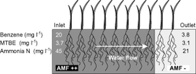
In March 2011 five samples of roots (each about 10 g) were taken from the ‘front’ (near the inlet) and five from the ‘rear’ (near the outlet; 10 samples in total) of the illustrated constructed wetland (5 m long, 1.15 m wide, 1.25 m deep; inflow rate 6 l h−1) planted with P. australis, which is being used in a compartment transfer experiment close to Leuna, Germany (Seeger et al., 2011). Parts of the sampled roots were stained with ink (Sheaffer, Middlesex, UK) and vinegar according to Vierheilig and colleagues (1998) to highlight AMF structures, and the degree of colonization by AMF was roughly estimated by inspecting the stained roots under a stereomicroscope and estimating approximate ratios of mycorrhizally colonized to non-colonized root lengths. Substantial degrees of AMF colonization were observed in all five root samples from the ‘front’ part of the wetland (40%, 25%, 25%, 60% and 80%). In contrast, no colonization of P. australis roots was observed in samples from the rear part, where there was no gravel substrate and the roots formed a dense root mat. These microscopic observations are consistent with results of nested PCR analysis of a 400 bp fragment of the nuclear large ribosomal subunit using the primer pairs LR1/FLR2 and FLR3/FLR4 (Gollotte et al., 2004) and Taq PCR Mastermix (Qiagen, Hilden, Germany). DNA extracted (using a DNeasy Plant Mini-Kit, Qiagen) from all samples from the front part of the wetland yielded fragments of expected size (for AMF), while DNA extracted from samples from the rear part yielded no PCR products. The concentrations of pollutants (benzene, methyl tert-butyl ether/MTBE and ammonia N) shown in the figure have been taken from Seeger and colleagues (2011).